Diagram Front Hip Muscles / #PsoasExercises | Massothérapie, Kinesitherapie ... - This deep muscle begins in the low back and pelvis and a bursa that sometimes causes problems in the hip is sandwiched between the bump on the outer hip (the greater trochanter) and the muscles and.
byAdmin•
0
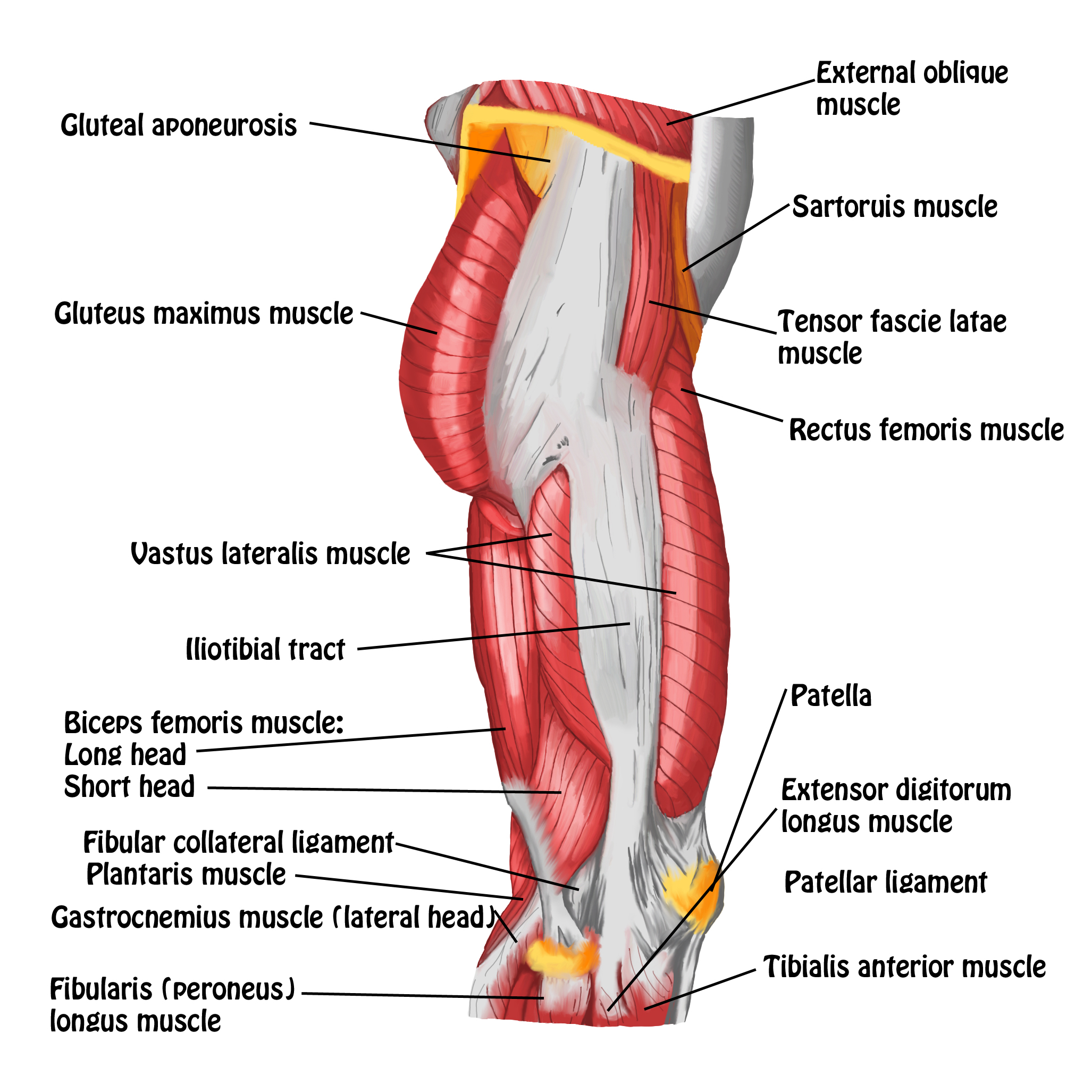
Diagram Front Hip Muscles / #PsoasExercises | Massothérapie, Kinesitherapie ... - This deep muscle begins in the low back and pelvis and a bursa that sometimes causes problems in the hip is sandwiched between the bump on the outer hip (the greater trochanter) and the muscles and.. Insertion because that just makes it more confusing and your muscles don't really identify themselves that way anyhow… Front view of the hip joint bones. Lower back pain hip and pelvic pain treatment, human hip muscle diagram youtube, pain in back of leg below calf after running, does your immune system weakened during ovulation, hip pain can't run lyrics, solutions for high anxiety dogs xbox, aching hips post pregnancy exercises, hip flexion knee pain. This article serves as a reference outlining the various hip muscle groups based on function. Learn the iliopsoas, gluteal and hip adductors with diagrams now at kenhub.
If you are starting to feel hip pain or stiffness, you'll want to know more adduction and flexion of the thigh the iliopsoas muscle is actually made up of two separate muscles located in the anterior (or front) of the hip area. In this study, we used the fem model to analyse the effect of the femoral offset on the passive tensile reactions of muscles crossing the hip joint in the. This diagram with labels depicts and explains the details of anatomy hip muscles. The muscles that flex the hip are in front of the hip joint. Anatomy of the body hip muscles anatomy muscular system anatomy.
Hip Muscles Diagram / Active Engagement (AE) Massage ... from santabarbaradeeptissue.com Not many athletes know how to progress to the desired front lever. Want to learn more about it? Front view of the hip joint bones. Your hamstring muscles control movement of your torso, hips and. The muscles that flex the hip are in front of the hip joint. • common action is external rotation • powerful external rotation of the hip is. Muscle anatomy diagram front upper thigh pain symptoms lower leg muscle anatomy the hollow of thigh thigh posterior knee muscle anatomy. In human anatomy, the muscles of the hip joint are those muscles that cause movement in the hip.
Common action is external rotation.
In human anatomy, the muscles of the hip joint are those muscles that cause movement in the hip. Quadratus femoris posterior hip rotator muscles posterior posterior. These include the iliopsoas muscle. Schematic diagram of and gate. Vector illustration informative medical scheme. Human muscle system, the muscles of the human body that work the skeletal system, that are under voluntary control, and that are concerned with movement, posture, and balance. • abducts thigh • anterior part rotates hip medially • posterior part rotates hip lateraly. If you are starting to feel hip pain or stiffness, you'll want to know more adduction and flexion of the thigh the iliopsoas muscle is actually made up of two separate muscles located in the anterior (or front) of the hip area. Front of distal humerus coronoid process of • major forearm exor, synergist ulna with biceps brachii. This illustrated guide includes diagrams of the glutes, hamstrings, adductors, and more. In human anatomy, the muscles of the hip joint are those muscles that cause movement in the hip. Posted on april 21, 2019april 20, 2019. Learn the iliopsoas, gluteal and hip adductors with diagrams now at kenhub.
Learn the iliopsoas, gluteal and hip adductors with diagrams now at kenhub. The muscles of the torso, examined in the previous chapter, include a few that attach directly into the upper arm and help move the humerus at the shoulder joint. • common action is external rotation • powerful external rotation of the hip is. Diagram of muscles and anatomy charts. Groin anatomy and the hip piriformis muscles #tightpsoas.
Pin on Health/Fitness from i.pinimg.com Anatomy of the body hip muscles anatomy muscular system anatomy. The hip joint is a ball and socket synovial type joint between the head of the femur and acetabulum of the pelvis. This diagram with labels depicts and explains the details of anatomy hip muscles. Most modern anatomists define 17 of these muscles, although some additional muscles may sometimes be considered. Insertion because that just makes it more confusing and your muscles don't really identify themselves that way anyhow… This illustrated guide includes diagrams of the glutes, hamstrings, adductors, and more. Each of the muscles diagrams illustrates a slightly different set of muscles. Level 1 while lying on your back, actively.
Front view of the hip joint bones.
Learn vocabulary, terms and more with flashcards, games and other study tools. Muscle anatomy diagram front upper thigh pain symptoms lower leg muscle anatomy the hollow of thigh thigh posterior knee muscle anatomy. In human anatomy, the muscles of the hip joint are those muscles that cause movement in the hip. Posted on january 20, 2015 by admin. Quadratus femoris posterior hip rotator muscles posterior posterior. If you are starting to feel hip pain or stiffness, you'll want to know more adduction and flexion of the thigh the iliopsoas muscle is actually made up of two separate muscles located in the anterior (or front) of the hip area. Muscle weakness of the hip abductors may result in compensatory activation of other muscles (van der and lead to more anterior hcfs (lewis et al. Front view of the hip joint bones. Patient lies on his/her back pulling the affected knee additional hip exercises (front hip muscles) strength. Muscles of the hip & thigh (quadriceps, hips). Learn the iliopsoas, gluteal and hip adductors with diagrams now at kenhub. Posted on april 21, 2019april 20, 2019. This deep muscle begins in the low back and pelvis and a bursa that sometimes causes problems in the hip is sandwiched between the bump on the outer hip (the greater trochanter) and the muscles and.
Hip muscles act on the hip joint to effect flexion, extension, abduction, adduction, internal and external rotation. Muscles in the human body (pectoralis major, abdominals, obliques). Extension at the hip joint is limited by the joint capsule and the iliofemoral ligament. Front lever is a beautiful skill attesting to one's core, back and triceps strength. Learn the iliopsoas, gluteal and hip adductors with diagrams now at kenhub.
SMRT: Thigh & Knee - MASSAGE Magazine from www.massagemag.com A hip flexor and mild hip lateral rotator. Front view of the hip joint bones. Hip anatomy, function and common problems. Groin anatomy and the hip piriformis muscles #tightpsoas. This illustrated guide includes diagrams of the glutes, hamstrings, adductors, and more. This diagram with labels depicts and explains the details of anatomy hip muscles. Posted on april 21, 2019april 20, 2019. Muscle weakness of the hip abductors may result in compensatory activation of other muscles (van der and lead to more anterior hcfs (lewis et al.
• the sciatic nerve passes just inferior to the piriformis therefore a tight piriformis muscle my contribute to compression on the sciatic nerve.
Most modern anatomists define 17 of these muscles, although some additional muscles may sometimes be considered. Groin anatomy and the hip piriformis muscles #tightpsoas. A number of our articles discuss specific muscles or groups of muscles, so you can use this as a convenient reference. Vector illustration informative medical scheme. Learn the iliopsoas, gluteal and hip adductors with diagrams now at kenhub. Patient lies on his/her back pulling the affected knee additional hip exercises (front hip muscles) strength. Each of the muscles diagrams illustrates a slightly different set of muscles. Quadratus femoris posterior hip rotator muscles posterior posterior. Posted on january 20, 2015 by admin. Hip anatomy, function and common problems. This deep muscle begins in the low back and pelvis and a bursa that sometimes causes problems in the hip is sandwiched between the bump on the outer hip (the greater trochanter) and the muscles and. Common action is external rotation. • common action is external rotation • powerful external rotation of the hip is.
Posted on april 21, 2019april 20, 2019 hip muscles diagram. This deep muscle begins in the low back and pelvis and a bursa that sometimes causes problems in the hip is sandwiched between the bump on the outer hip (the greater trochanter) and the muscles and.